Passive Knee Extension Test of Hamstring Extensibility
Contents
Preamble: A ROM Protocol for Passive Knee Extension
Positioning
Anatomical Referencing and Gravity Correction
Exercise Set-up: Range of Movement and Return Force
Data Acquisition and Reduction
References
Acknowledgements
Preamble: A ROM Protocol for Passive Knee Extension
The Range of Motion or ROM protocol is a relatively new option on the KIN-COM dynamometer. Its unique feature is the ability to expand the range of movement by up to 20 degrees beyond the stop angle. It is sometimes termed a soft-tissue stretching protocol because of this feature. It may be considered to be a variation on an isotonic exercise mode in that it is a force sensitive mode. The achieved range of motion is dependent on the force elicited during the movement. That is, the full range of movement may not be completed if the preset threshold (Return) force is elicited during the movement.
The maximum velocity of movement is limited in this protocol. This results in a smooth (typically slow) movement at constant angular velocity through range until the force threshold is matched. The movement is then stopped and reversed in direction back toward the start angle. In our variation to this protocol, the lever arm is held for 1 or 3 seconds at the angle at which the Return Force is elicited.
We implemented this protocol in 1996 as a passive knee extension to determine hamstring extensibility. Poor hamstring flexibility has been postulated as a factor in the development of hamstring injury (Sutton 1984). Both accurate measurement techniques and normal ranges of hamstring length must be established prior to undertaking studies to determine any correlation with predisposition to hamstring injury (Gajdosik et al 1993). Accurate measurement procedures are therefore essential to evaluate the effect of intervention programs and stretching regimen.
The passive straight leg raise and passive and active knee extension tests have been commonly used as clinical tests of hamstring muscle length. The active knee extension (AKE) test has been proposed to be a more objective measure of hamstring tightness than the straight leg raise for a variety of reasons. Gajdosik and Lusin (1983) produced reliability correlation coefficients of 0.99 for each tested extremity when performing test-retest measurements in an AKE test, with knee extension angle as the dependent variable. The authors attributed the high reliability to limitation of pelvic motion and precise instrument placement. Whilst admitting motion of the pelvis was not eliminated, they suggested that pelvic motion was reduced in comparison to the straight leg raise testing procedure. Sutton (1984) also emphasised the need for adequate stabilisation of adjacent articulations to allow movement to occur at only one of the two joints which the hamstrings affect and then advocated the use of the knee extension test for this purpose. The reasoning for this recommendation was not only to provide improved stabilisation and hence accuracy, but also in an attempt to simulate conditions causing hamstring strains.
It is necessary to have a test that produces reliable and accurate measurements of hamstrings extensibility. Although variations on the passive knee extension (PKE) test have been previously used clinically and in research (Magnusson et al., 1995; McHugh et al., 1992; Starring et al., 1988), its reliability has not been adequately investigated. If adequate stabilisation of adjacent joints can be achieved, and the PKE test is shown to be reliable from limb to limb and day to day then the extensibility of the hamstring muscle group can be accurately assessed for comparison of injured and uninjured limbs, for prediction of injury, for objectively measuring treatment outcomes and for determining return to sport after rehabilitation.
Positioning
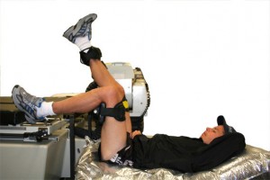
A special supporting bench is used to position the subject. This bench is lower than the Kin-Com bench and allows the subject to be positioned with their knee joint in line with the axis of rotation of the dynamometer and the hip joint immediately below the knee joint (a plumb bob is used for the vertical alignment of the knee and hip joints. The knee joint centre (lateral femoral condyle) is aligned with the axis of rotation of the dynamometer by moving the supporting bench and the dynamometer head. The thigh is stabilised with the Universal stabiliser attachment and the trunk is horizontal on the supporting bench. Thus, the hip is positioned in 90 degrees of flexion. The other lower extremity is also flexed at the hip and knee.
A trunk stabilising bag is used to minimise trunk and head movement. This bag is filled with polystyrene balls and a valve is fitted at one end. When the subject has been positioned in the bag with a supported horizontal trunk and head, with their arms folded across their chest, straps are placed around the bag and the subject. The air in the bag is then removed by a suction pump. The bag is moulded to the subject as it is evacuated, and the subject is restrained in the form of the bag.
A special supporting bench is used to position the subject. This bench is lower than the Kin-Com bench and allows the subject to be positioned with their knee joint in line with the axis of rotation of the dynamometer and the hip joint immediately below the knee joint (a plumb bob is used for the vertical alignment of the knee and hip joints. The knee joint centre (lateral femoral condyle) is aligned with the axis of rotation of the dynamometer by moving the supporting bench and the dynamometer head. The thigh is stabilised with the Universal stabiliser attachment and the trunk is horizontal on the supporting bench. Thus, the hip is positioned in 90 degrees of flexion. The other lower extremity is also flexed at the hip and knee.
In early studies a trunk stabilising bag was used to minimise trunk and head movement. This bag is filled with polystyrene balls and a valve is fitted at one end. When the subject has been positioned in the bag with a supported horizontal trunk and head, with their arms folded across their chest, straps are placed around the bag and the subject. The air in the bag is then removed by a suction pump. The bag is moulded to the subject as air is evacuated, and the subject is restrained in the form of the bag (Figure 1).
Figure 1.
More recently, the thigh support system has been modified to provide greater stability. The trunk stabilising bag has been removed and a layer of high density foam padding on the supporting bench has been used to support the pelvis, trunk and head (Figure 2). Pelvic tilt (flattening of the lumbar spine) is monitored using a Chattanooga Pressure Biofeedback unit (Figure 3) from which the pressure signal is captured in a separate data acquisition system. The resistance pad is located distally on the leg (with the furthest edge of the pad about two centimetres proximal to the anterior ankle crease). It is important that the centre of the resistance pad is aligned with a whole centimetre value on the lever arm ruler – this number is then the lever arm length.
The resistance pad is located distally on the leg (with the furthest edge of the pad about two centimetres proximal to the anterior ankle crease). It is important that the centre of the resistance pad is aligned with a whole centimetre value on the lever arm ruler – this number is then the lever arm length.
Anatomical Referencing and Gravity Correction
Anatomical referencing is performed by positioning the leg at 90 degrees to the thigh ie. the leg is positioned horizontally as the thigh has previously been positioned vertically. This position is essentially a 90 degree knee flexion determined by goniometry. A positive direction is defined in the direction of knee extension.
Gravity correction or gravity compensation is performed with the leg in the same position. The full leg and foot weight should be registered by the load cell as the hamstrings are relaxed and the quadriceps are not on stretch. Check that the subject has relaxed the leg and foot by gently moving the foot. Accept and make note of the limb weight reading once it is steady.
Exercise Set-up: Range of Movement and Return Force
To determine the stop angle the lever arm was moved beyond full knee extension and then the subject was instructed to actively extend the knee and hold the position. The lever arm is then moved in the direction of knee flexion until it is in contact with the actively extended leg. This (active knee extension) position is defined as the stop angle. The start angle is then set at either 90 120, or 135 degrees of knee extension. Once both start and stop angles are set, the ROM protocol requires the lever arm to be moved to the stop angle. At this angle the resistance force is assessed by the Kin-Com. This is accepted as the Return Force. The Maximum Force must then be set at approximately 50 N more than the Return Force. The present protocol then changes the Return Force to twice the limb weight noted previously. The protocol then requires the Maximum Force to be reset. The amount of extended ROM (beyond the stop angle) may then be set at 20 degrees.
For the limb to hold the limb at the Return Angle, an alteration to the Setup is required. The Minimum Force value in the isometric settings within the force limits options is changed to -200N and the hold time is changed to 1 or 3 seconds, as preferred. The ‘Relax after Contraction’ setting is changed to NO.
A feature of our implementation of the ROM protocol is that the resistance force is maintained at the constant level of twice leg weight for speeds of movement below 5 degrees per second. Once positioned, the subject are asked to relax and not to move any other part of their body while their knee is extended and flexed passively by the Kin-Com. The knee is cyclically extended and flexed 30 times. The subject remains in position on the stabilising bag between testing of the left and right legs. The angle which the Kin-Com reached when achieving (but not exceeding) the constant force level, is displayed on the Kin-Com computer screen for each repetition (and can be stored in the .CPM datafile), and is recorded as the PKE angle. This absolute PKE angle is the dependent variable. PKE angle is an angle in whole degrees (no decimal places – if anyone with programming influence at Chattecx Corporation reads this and can increase the resolution of the angle measurement to display to one decimal place, this would really be valuable!)
The parameters for the PKE Test are listed below.
ROM Protocol Settings – Exercise Mode
Control of Constant SPEED Isokinetic Forth 5 deg/s Back 10 deg/s
Motion and Initiating Force Con/Ecc Forth 0 N Back 0 N
Force Limits: Minimum 0 N Maximum 200 N
Return Force 2 x Leg Wt. Pause 1s at Stop
Relax after Pause No
Turn Points: Acceleration Settings Low Deceleration Setting Low
ROM Expansion 20 degrees Exercise Time 600s
Data Acquisition and Reduction
Visual inspection of the data suggested that a plateau in the angle of knee extension was not clearly evident over the 30 repetitions. The data was therefore averaged over five trials to produce six data sets. A three factor (testing occasion x limb x trial) analysis of variance with repeated measures performed on this data did not demonstrate any significant three-way or two-way interactions.
On selection of the average of trials 21 to 25, the measurement of PKE angle was shown to be reliable from day to day in normal subjects with no significant difference between sides. The results of this study indicate that there is a creep response in extensibility of PKE. Based on regression analysis, a plateau in PKE angle was not established prior to 30 repetitions. Further research with greater than 30 trials is recommended to establish if and when a plateau exists using this stretching protocol.
References
Ainslie TR and Beard DJ (1996) Quantification of quadriceps passive resistance using isokinetic dynamometry. Physiotherapy 82(11): 628-630.
Gajdosik RL and Lusin G (1983) Hamstring muscle tightness reliability of an active knee extension test. Physical Therapy 63: 1085-1090.
Gajdosik RL, Rieck MA, Sullivan DK and Wightman SE (1993) Comparison of four clinical tests for assessing hamstring muscle length. Journal Orthopedic of Sports and Physical Therapy 18: 614-618.
Magnusson SP, Simonson EB, Aagaard P, Gleim GW, McHugh MP and Kjaer M (1995) Viscoelastic response to repeated static stretching in the human hamstring muscle. Scandinavian Journal of Medicine & Science in Sports 5: 342-347.
McHugh MP, Magnusson SP, Gleim GW and Nicholas JA (1992) Viscoelastic stress relaxation in human skeletal muscle. Medicine and Science in Sports and Exercise 24: 1375-1382.
Starring DT, Gossman MR, Nicholson GG, Lemons J (1988) Comparison of cyclic and sustained passive stretching using a mechanical device to increase resting length of hamstring muscles. Physical Therapy 68: 314-320.
Sutton G (1984) Hamstrung by hamstring strains: A review of the literature. Journal of Orthopaedic and Sports Physical Therapy 5: 184-195.
Taylor DC, Seaber AV and Garrett WE (1990) Viscoelastic properties of muscle-tendon units: The biomechanical effects of stretching. American Journal of Sports Medicine 18: 300-309.
Acknowledgements
Peter Hamer and I first started talking about using the Kin-Com with the subject positioned on a supporting bench in about 1992. More recently, I have used a variation on this protocol for testing hamstring extensibility in elite Australian Football League players. Liza Devine, Michal Lashkar and Tambu Masaya then completed a research project on “The reliability of a Passive Knee Extension Test” as part of the Postgraduate Diploma in Sports Physiotherapy at the School of Physiotherapy at Curtin University of Technology.
[Top]
